| Schema of the fully automated cell segmentation method. |
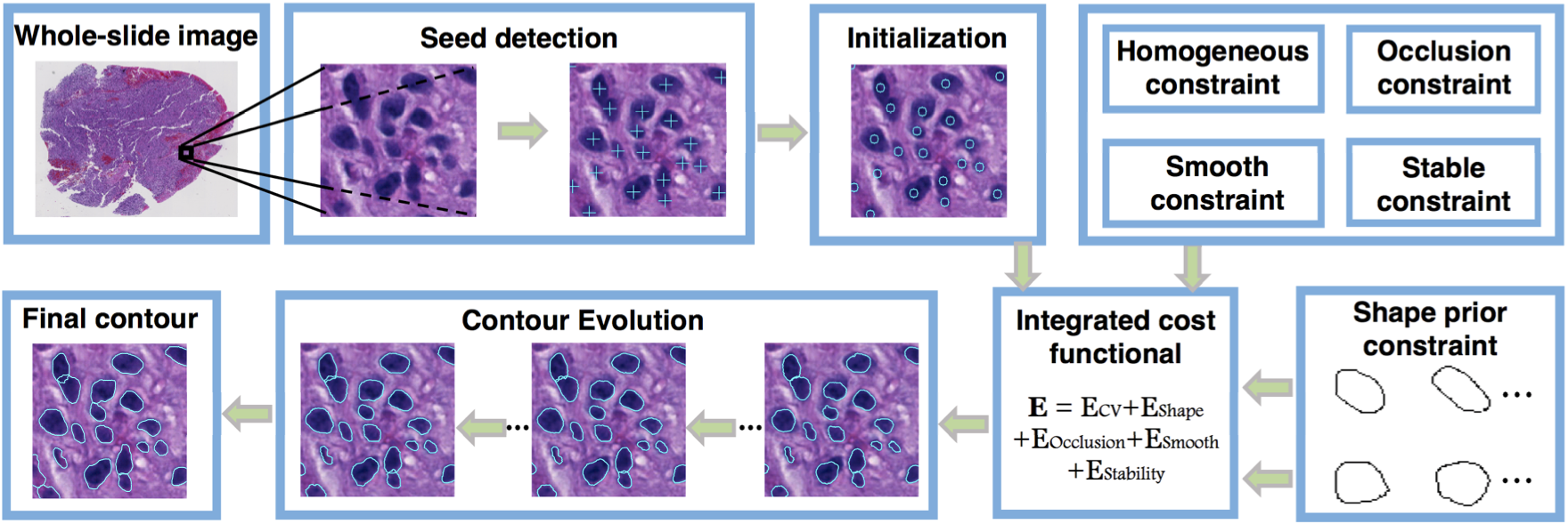 |
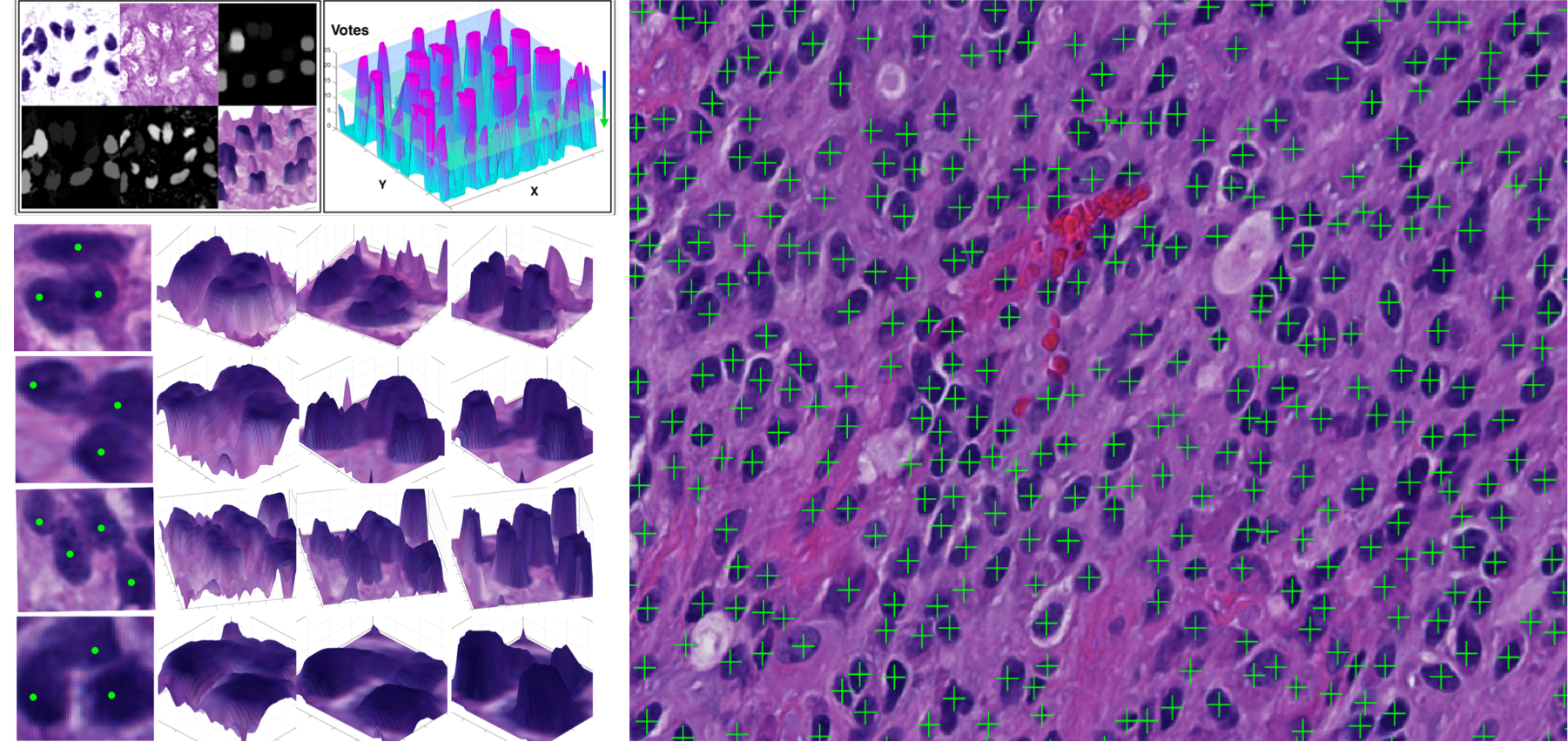
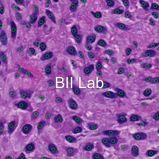
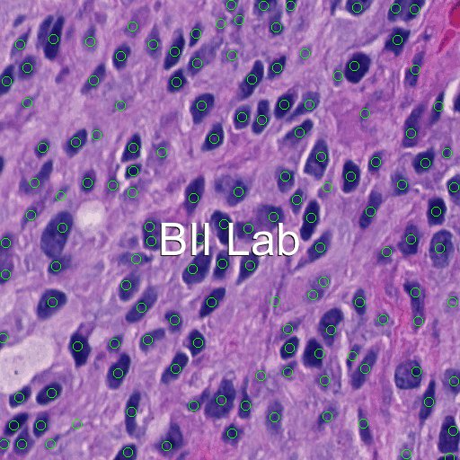
| The cell seed detection algorithm utilizes joint information of spatial connectivity, distance constraint, image edge map, and a shape-based voting map derived from eigenvalue analysis of Hessian matrix across multiple scales. |
 |
| Variational level set method with a new shape prior term, an adaptive contour occlusion penalty term, and a boundary attraction term. |
 |
|
 |
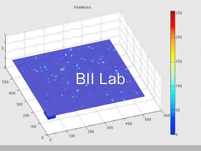
| we have applied our 2D method to a randomly selected 2D image slice of a 3D testing fluorescent imaging volume. |
 |
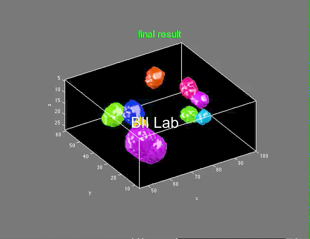
| The ex-vivo 3D fluorescent cell segmentation results: |
 |
|
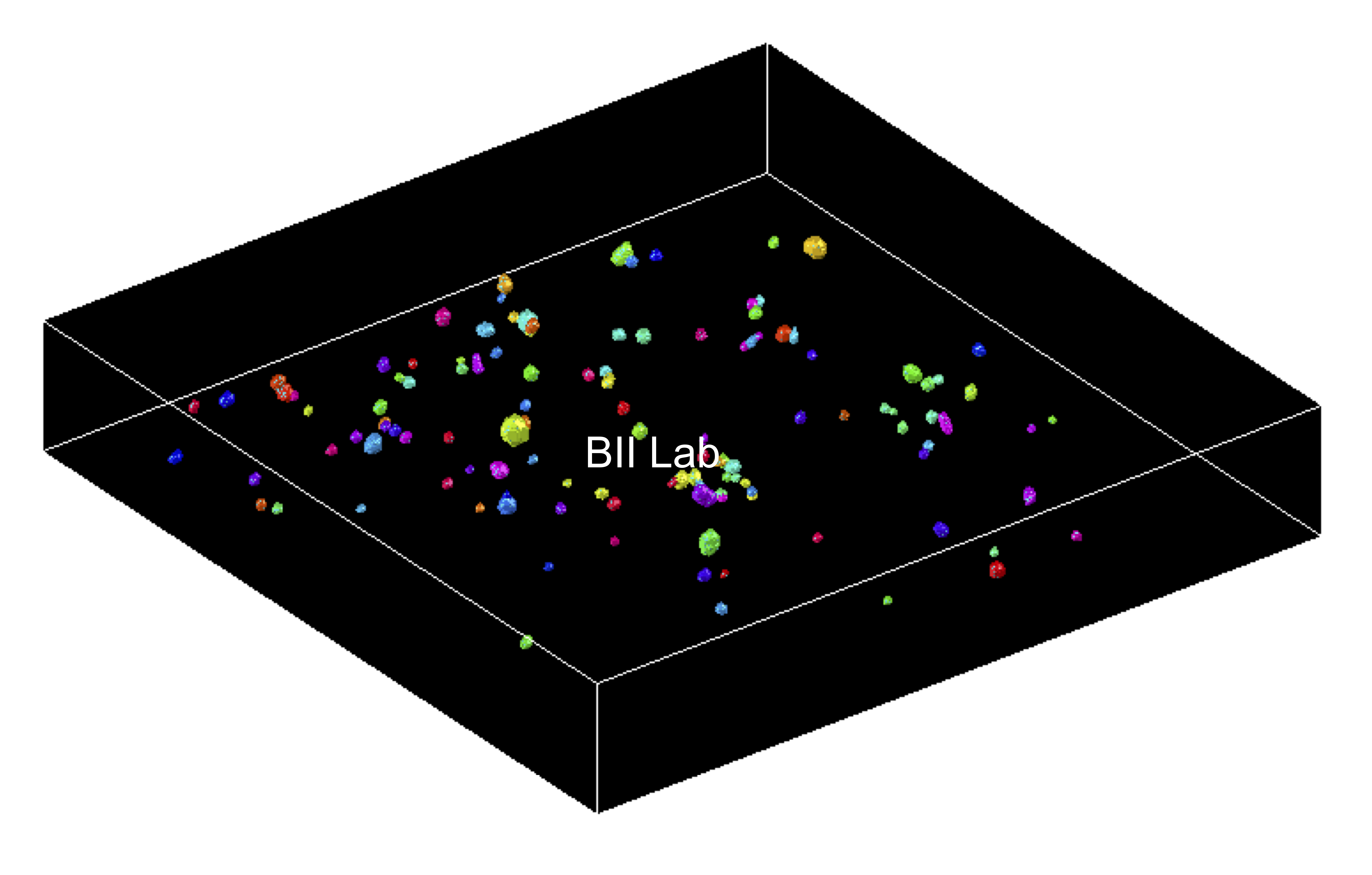 |
| 3D cell surfaces are projected to 2D image planes for qualitative cell segmentation assessment: |
 |
| 3D segmented cells are overlaid with a 2D image plane for qualitative cell segmentation assessment: |
 |
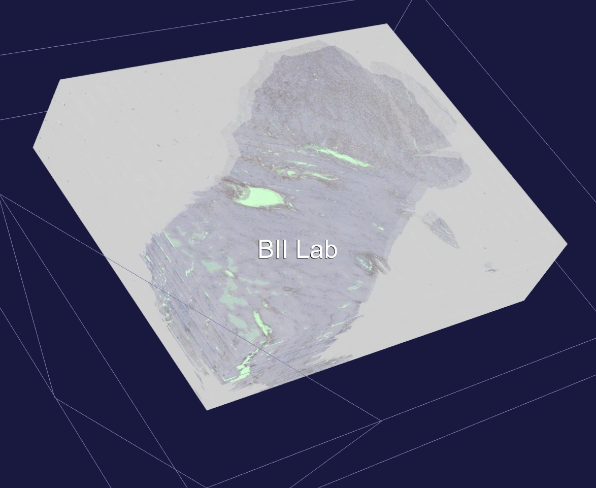
| 3D Hepatic artery segmentation. |
 |